PFNTS - Method

Pulsed Fast Neutron Transmission Spectroscopy (PFNTS) is an imaging method that exploits the characteristic cross-section structure (resonances) of different isotopes in the energy-range En=1-10 MeV (see in fig.1). The method holds promise for identifying and detecting a broad range of explosives due to its ability to determine simultaneously the identity and density distribution of the principal elements present in explosives, such as C, O and N.

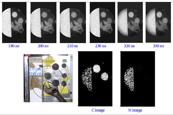
Since 2004 PTB and Soreq NRC, in collaboration with the Weizmann Institute of Science are developing a high spatial resolution (sub-mm) PFNTS system based on a time-resolved integrative optical neutron (TRION) detector.

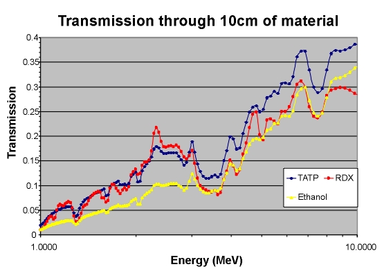
In PFNTS the inspected object is irradiated with neutrons of a broad spectral distribution in the above energy range. If the inspected object contains elements that exhibit sharp cross-section resonances, the transmission neutron spectrum will be modified such that it will exhibit dips and peaks at specific energies corresponding to these resonances.

Thus, the transmission spectrum carries information about the elemental composition of the object. Fig. 2 shows a calculated transmission through 10 cm thick objects comprising TATP (an improvised explosive), RDX (a military explosive) and ethanol which all have similar densities. Indeed, these three items would look the same to an X-ray probe, which measures density only, whereas PFNTS determines the characteristic stoichiometric ratio of elements in the substance.
  TOF
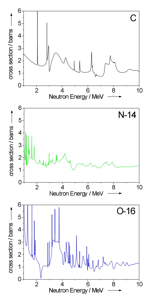
Fig.1 Cross sections of C, N, O in the MeV region.
Thus, the neutron transmission spectrum can be used to distinguish between explosives and benign materials of similar physical density.

A pre-requisite for PFNTS is imaging at several well defined neutron energies. This is achieved by neutron Time-of-Flight (TOF) spectroscopy, requiring pulsed neutron beams and imaging systems with capability for TOF measurements.
TOF
Fig.2 calculated transmission through 10 cm thick objects comprising
TATP (an improvised explosive), water and polyethylene, which all
have similar density.

As an example of the PFNTS technique Fig. 3 shows an object, consisting of melamine (which simulates here a commercial, nitrogen-rich explosive), several carbon rods and a steel wrench, that was consecutively imaged at various neutron energies. The top row of Fig 3 shows the neutron transmission images, marked with the corresponding TOF windows. At first glance they all look identical; however, with appropriate image arithmetic, using the characteristic cross section structure of the elements the net carbon and nitrogen distribution of the objects can be derived (Fig 3, lower right). Also, as expected, the steel wrench disappears completely in this process. TOF
Fig.3 Neutron radiographs of melamine block, carbon rods and steel wrench in 6 TOF - bins. Lower row shows carbon and nitrogen distribution in sample, based on analysis of neutron resonances.